PROJECT FUNDED BY:
3D bioprinted liver tissue with vascular system as an innovative model for assessing drug toxicity and effectiveness of oncological therapies, project financed by the European Union from the European Regional Development Fund, Operational Programme Smart Growth 2014–2020. The project is implemented under the National Centre for Research and Development competition 3/1.1.1/2020 Fast Track 3_2020. Implementation period: 2021–2023

R&D project coordinator
Assoc. Prof. Michał Wszoła MD, PhD
Project funded by:
Time frame:
Project leader:
Scientific and industrial consortium made up of the Foundation of Research and Science Development and Polbionica sp. z o.o.
3D BIOPRINTED LIVER TISSUE WITH VASCULAR SYSTEM AS AN INNOVATIVE MODEL FOR ASSESSING DRUG TOXICITY AND EFFECTIVENESS OF ONCOLOGICAL THERAPIES
PROJECT OBJECTIVE:
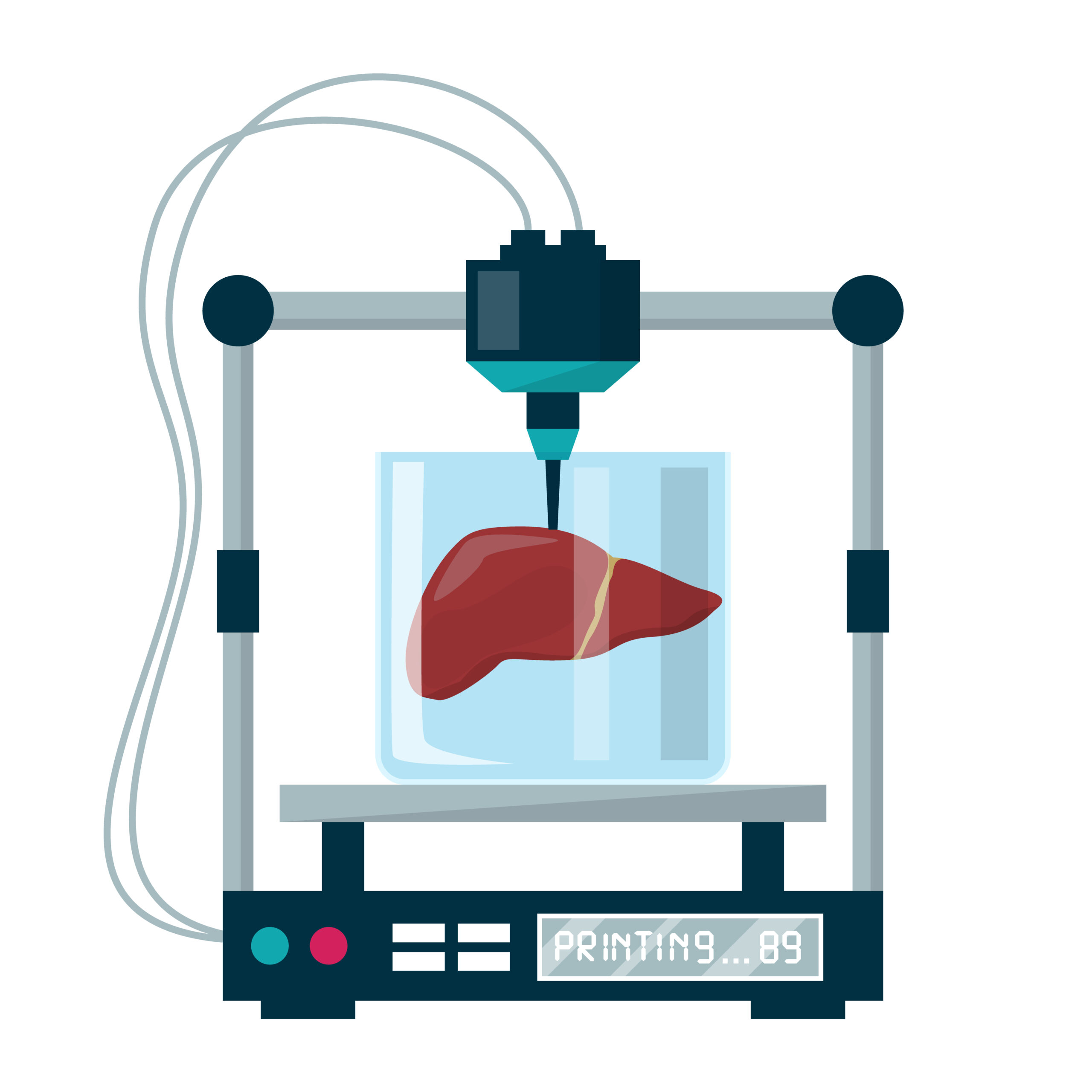
The aim of the project is to develop innovative bioinks dedicated to the bioprinting of a 3D model of bionic liver tissue with vascular system for toxicological tests and a 3D model of bionic liver tissue with vascular system and neoplastic foci for testing the effectiveness of therapeutic substances.
The idea of creating a 3D model of bionic liver tissue with vascular system is an answer to the imperfections of current cell culture models which—due to their two- or three-dimensionality—can give toxicity or substance effectiveness assessment results that are difficult to transfer to a living organism in subsequent stages of research, i.e., to the stage of preclinical trials. Using the proposed 3D models, including models with neoplastic foci, will provide a preliminary estimate of the optimal variant and concentration of the test substance.
This solution, innovative on a global scale, will find application in toxicological studies and effectiveness tests of new therapeutic substances, which will facilitate a significant reduction or perhaps even a complete withdrawal from preclinical research with the participation of animals in the future in favour of equally effective tests with the use of 3D bioprinted models with a vascular system.

WHAT ARE BIOINKS?
IMPORTANT INFORMATION
The composition of the bioprinting ink for a 3D model of bionic liver tissue with vascular system should provide mechanical strength to the organ, enable cell adhesion, and direct the cells for further growth and development. The use of this bioink should ensure: high cell survival (at least 80%), proper functioning of the tissue metabolic system (e.g., albumin and heparin production), and stability and durability of the model. An important feature of biomaterials for the bioprinting of research systems is their adequate translucency for observation under a microscope.
The creation and use of a 3D bionic liver tissue model at the stage preceding preclinical trials will enable advanced in-vitro studies and facilitate optimization of preclinical trials and selection of optimal variants and concentrations of therapeutic substances. The 3D bioprinting technology of tissue systems will find applications in the pharmaceutical industry for testing new drugs and will constitute an alternative to animal testing, providing equally reliable test results.
The main recipients of research on 3D bioprinted tissue models will be pharmaceutical companies interested in expanding the scope of research in the process of therapeutic substance development.
Bioinks in the form of a finished product will be offered to research centres and entities in the biotechnology industry.
The initiator of the project is an eminent surgeon, transplantologist Michał Wszoła, MD, PhD. Together with a skilled research and management team, they constitute a guarantee of the research success of this venture.


